Posts Tagged ‘osteoarthritis’

When your client says, “But my MRI says”, then you say……

Friday, April 15th, 2016

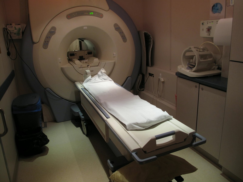
Magnetic resonance imaging (MRI) is one of the most sensitive diagnostics currently available. It has frequently been the “last word” on pain, surgery and recommended limitations on activity. But should your client really never lunge or squat again because their doctor took an MRI and it showed some pathological condition?

Consider this review I did of a couple of studies on the matter. You may change the conversation you have with your clients once finished reading this.

Guermazi et. al. (2012) used magnetic resonance imaging to look at knees where radiographic imaging (x-rays) showed no osteoarthritic (OA) changes. OA is generally diagnosed through examination and x-ray. X-rays can identify bony changes to the joint but they cannot identify soft tissue pathologies. The purpose was to use the more sensitive MRI to detect structural lesions associated with OA and their relationship to age, sex and obesity.
MRI
710 subjects age 50 or older participated in the study (mean age 62.3 years). Out of the 710 subjects, 206 (29%) had painful knees.

Overall, 610 (89%) of the subjects showed some abnormality of the knee. Three most common findings of abnormalities in the knee were osteophytes, cartilage damage and bone marrow lesions. These abnormalities increased with age.

The study concluded that 91% of those who did have pain in their knee also had abnormal MRI’s, leaving 9% of those with painful knees having normal MRI’s. And 88% of those with no pain in their knees showed abnormalities in the MRI. The authors also noted that those with the highest amount of abnormalities in their MRI were those identified with mild pain and not those with moderate or severe pain (emphasis mine).

Another study in European Spine Journal (Kato et al. 2012) looked at MRI’s of the cervical spine of 1211 asymptomatic patients. The subjects were both men and women equally distributed between the ages of 20 years to 70 years. All of the subjects had both an MRI and neurological exam by a spinal surgeon.

Findings from the MRI of spinal cord compression, spinal cord signal changes and disc compression were noted. Increased signals on an MRI are associated with an abnormal state of the tissue such as scarring of inflammation.

For a disc bulge to be considered pathological it had to measure more than 1 millimeter from the vertebral body.
cervical spine degeneration
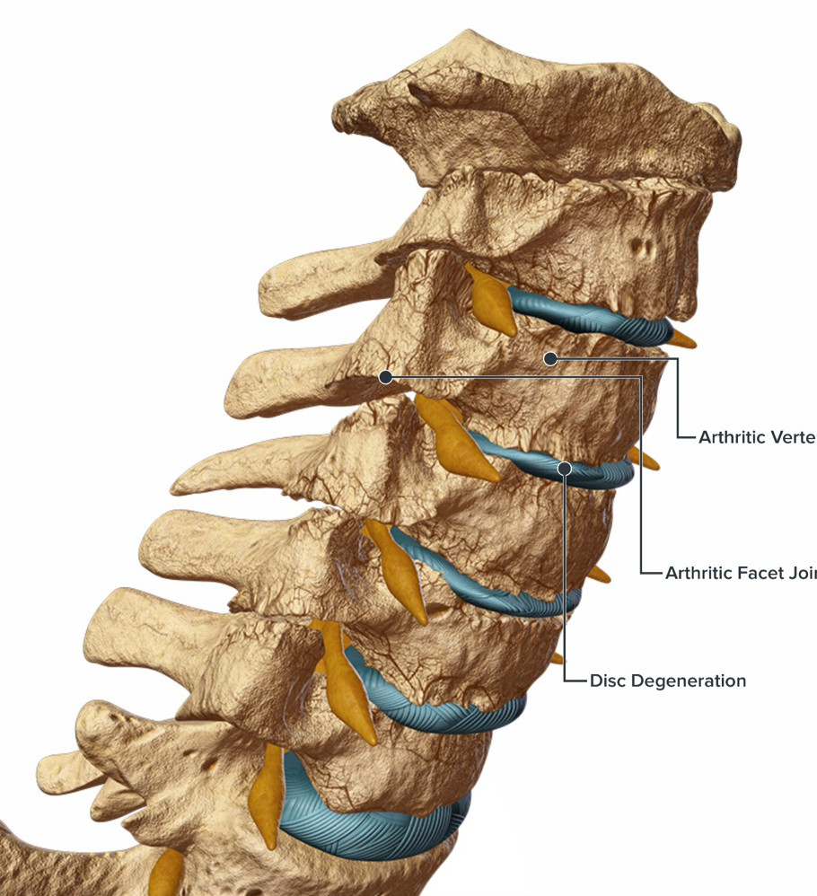
Of the 1211 asymptomatic subjects studied, 64 (5.3%) had spinal cord compression. High intensity signal changes were seen in 28 (2.3%) and disc bulging was seen in 1061 (87.6%) of subjects. Prevalence of these findings was significantly higher in people over 40 years of age.

If we consider the findings of both these studies, it is now clear that degenerative changes to the body are a normal part of aging and do not directly correlate with pain. Clients may experience stress or fear when learning of abnormalities in any joint or soft tissue following imaging studies done on them. Even if they are not in pain but have experienced pain in the past, the knowledge of degenerative changes are often communicated by medical professionals and perceived by individuals as the sole source of their pain. These studies clearly demonstrate that an individual can have many abnormal finding in the neck and knees and have no pain.

Clients who believe that the degenerative changes on their imaging will lead to pain may potentially act with self-limiting and guarded movements as well as an expectation of pain. This has the potential to decrease their functional capacity, increase anxiety about certain exercises or activities and view surgery as a necessary step to resolution.

Although I’ve suggested that your conversation should change with your clients, when you understand what these studies (and others) are telling us, we must remember that your client’s paradigm may not easily change. Their beliefs may be entrenched in an outdated pain/imaging relationship, especially if their doctor leads them to believe that the MRI finding is the final word.

They need proof. And ultimately that proof is movement confidence.

Guermazi, Ali August 2012. Prevalence of abnormalities in knees detected by MRI in adults without knee osteoarthritis: population based observational study (Framingham Osteoarthritis Study). BMJ, 345:e5339 doi: 10.1136/bmj.e5339).

Kato, Fumihiko et al. February 2012. Normal morphology, age-related changes and abnormal findings of the cervical spine. Part II: magnetic resonance imaging of over 1,200 asymptomatic subjects. Eur Spine J, DOI 10.1007/s00586-012-2176-4.

Does being overweight cause osteoarthritis?

Wednesday, June 9th, 2010

Lower back pain is only second to the common cold for missed works days in the United States. Eighty-five percent of people in the industrialized world will have an episode of debilitating back pain in their life. Many of these sufferers have symptoms related to osteoarthritis of the spine. According to the Arthritis Foundation, forty six million people in the United States are affected by osteoarthritis (OA).

The Arthritis Foundation defines OA as:
Osteoarthritis, a degenerative joint disease in which the cartilage that covers the ends of bones in the joint deteriorates, causing pain and loss of movement as bone begins to rub against bone. It is the most prevalent form of arthritis.

The hips, knees and shoulders are also common areas of OA along with the spine. When these joints are painful they will interfere with many exercise programs. Since the hips and knees are load bearing joints for walking, standing, etc., they assume a larger percentage of the impact forces during any upright activity. Reviewing any of the literature on osteoarthritis (OA), you will inevitably see being overweight and obesity commonly listed as “causes” of OA.

From a biomechanical standpoint, I do not believe that excess body weight causes OA. I do believe additional body weight can magnify other mechanical factors leading to OA. If we think about the cartilage as wearing out on the bones, the misconception is that it is the entire bone. Instead it is often an area on the bone that is subject to excessive friction relative to the other parts. I often describe this to my clients as a “hot spot” in the joint. If you apply more pressure to that spot (i.e. more body weight), it will certainly be more sensitive than if less weight were applied. Reducing a person’s body weight does reduce the pressure on the hot spot, but it does not reduce the hot spot itself.

The overweight person often says “I can’t exercise because it hurts”. We say, “It hurts because you don’t exercise”. The caveat here is first defining the type of exercise we are referring to. Far too many people who don’t exercise lump all forms of exercise into one big category. And if you speak to any fitness professional, we can break exercise into multiple categories. These can include but are not limited to:

• Corrective exercise
• Flexibility exercise
• Mobility exercise
• Restorative exercise
• Strength training
• Aerobic conditioning
• Metabolic conditioning
• Sport specific exercise
• Mind/body exercise

For us, proper exercise would refer to a program that first influenced how the body moves. This would incorporate a corrective exercise program that improved joint mechanics to better distribute the forces in the joint. This also does not take away the hot spot but it does reduce the pressure on it and improves the overall function of the joint. This in turn reduces the pain and slows the progression of further degeneration.

Let’s use the knees for example. In our example the person has genu valgum (“knocked knees”). This may in part be congenital. But it is also a result of muscle and soft issue influences on the knee joints. The valgus position of the knee increases the pressure on the outer knee and decreases pressure on the inner knee. Therefore, the forces that should be shared on the inner and outer knee are much greater on the outer knee. This often results in accelerated degeneration to the inner aspect of the knee while the outer aspect remains normal or closer to normal.
valgus knee stress
(Illustration credit: Ajit Chaudhari, PhD, Assistant Professor, Dept. of Orthopaedics Ohio State University)

Common imbalances associated with genu valgum are tightness of the hip ADDuctors and iliotibial band. In addition, the hip ABductors are weak relative to the adductors and do not resist the pull of the femur toward the midline of the body which leads to the valgus position of the knee. In many cases the foot and ankle are involved as well.

Addressing the body’s movement patterns through the muscles and connective tissue you improve the joint mechanics and decrease the stresses that precede and/or perpetuate the degeneration. Both sides of the knee joint now “share” the forces a little more thus removing some pressure from the “hot spot”.

This will allow the overweight individual to get all the other benefits associated with the exercise experience including:

• strength
• increasing energy
• improving sleep
• controlling weight
• decreasing depression
• improving self-esteem.
• combating osteoporosis
• reducing the risk of heart disease

Osteoarthritis is a degenerative process and the result of long term wear and tear. A strategy of ice and medications only douses the fire but never touches the fuel. And as long as you are moving the way you have always moved, the fuel will continue to build up. Changing the way you move through corrective exercises can cut off the fuel line to that fire.

See how Function First’s corrective exercise program can help you by clicking here.