Posts Tagged ‘neck pain’

Reverse Floor Block-Anti Keyboard Corrective Exercise

Tuesday, August 23rd, 2022

A client favorite, this exercise has multiple benefits to the upper extremities, thoracic spine and neck region. Utilizing the arm positioning and the cueing of the radial-ulnar and gleno-humeral joints, the motor system can access the peri scapular muscles in a very efficient way.

Lengthening through the chronically shortened muscles of the wrist and forearm and isometrically contracting the middle trapezius and rhomboids produces a noticeable change in the tissue tension and posture of the upper body.

Neck pain from your eyes?

Wednesday, April 27th, 2016

Chronic pain is complex, resulting from many inputs processed through the nervous system and the brain. As humans, we rely heavily on our vision to assess and navigate our environment and maintain balance.

Visual references are also one type of input the brain relies on to determine a potential threat to the organism. For example, have you ever found a bruise on your body that did not hurt until you noticed it there?

For those suffering from chronic neck pain, vision provides a great deal of feedback about cervical range of motion along with the mechano-receptors in the joints and soft tissue. The endpoint a person sees when turning his or her head and experiencing pain combines with a cluster of other information occurring at the same time to form the neuro-representation of the pain experience in the brain, or what Melzack (2001) calls a “neuro-signature.”

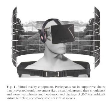
Harvie et al. (2015) investigated the role of visual feedback on neck pain. The researchers used a virtual-reality apparatus to alter the visual proprioceptive feedback that subjects received during cervical rotation. Subjects were seated with their torsos fixed to avoid contributing motion from the thoracic spine during cervical rotation. Twenty-four subjects with chronic neck pain were assessed for the onset of pain during cervical rotation to the left and right. They were asked to stop when they felt pain and to rate it on a scale of 0-10 at the point in the rotation where pain occurred. Each subject was then fitted with a virtual-reality headset that provided six different visual scenes for six trials. The image below is taken directly from the study by Harvie et al. (2015) and shows an illustration of the set up.

bogus vision article

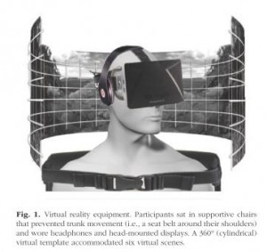
Researchers manipulated the virtual-reality scenes so that the visual cues did not match the actual cervical-rotation distance that subjects achieved on all trials. The virtual rotation provided by the headsets was either:

• 20% more than the actual rotation
• the same as the actual rotation
• 20% less than the actual rotation

This bogus visual feedback of plus or minus 20% made the subjects perceive that they were rotating their cervical spines 20% more or less than they actually were.

The results showed that when rotation was understated (subjects perceived their rotation was less than it actually was), pain-free range of motion increased by 6%. When rotation was overstated (subjects perceived their rotation was more than it actually was), pain-free range of motion decreased by 7%.

This study provides additional evidence to support the findings that pain is not generated solely from tissue damage. The bio-pyscho-social model acknowledges multiple inputs contributing to the pain experience.

Vision is one of many contributing inputs that the brain processes when assessing a threat to the body and therefore produces pain. The association of a specific neck range of motion identified visually, coupled with information from the motor system and proprioceptive system, creates a confirmed reference for past pain experiences. In other words, we’ve always had pain with this set of circumstances (neuro signature of matched proprioception, motor function, vision, vestibular), so we are supposed to have now. Hello pain.

It is plausible that visual input can also influence pain in other areas. For example, if a client has lower-back pain, forward flexion of the spine will bring her eyes closer to the floor, possibly presenting a painful or pain-free experience, depending on the client.

When designing a corrective program for clients where you believe the visual field is a factor, you could vary the visual field to minimize the visual association related to painful movements. Or you could keep the head still and create the motion you want from the bottom up-creating relative movement of the cervical spine in relation to the thoracic spine.

Interested in learning more about how we, at Function First and the Pain-Free Movement Specialists work with the chronic pain population? Enrollment is available only until April 29th here.

Melzack, R. 2001. Pain and the neuromatrix in the brain. Journal of Dental Education 65(12), 1378-82.

Harvie, D.S., et al. 2015. Bogus visual feedback alters onset of movement-evoked pain in people with neck pain. Psychological Science. doi:10.1177/0956797614563339.

When your client says, “But my MRI says”, then you say……

Friday, April 15th, 2016

Magnetic resonance imaging (MRI) is one of the most sensitive diagnostics currently available. It has frequently been the “last word” on pain, surgery and recommended limitations on activity. But should your client really never lunge or squat again because their doctor took an MRI and it showed some pathological condition?

Consider this review I did of a couple of studies on the matter. You may change the conversation you have with your clients once finished reading this.

Guermazi et. al. (2012) used magnetic resonance imaging to look at knees where radiographic imaging (x-rays) showed no osteoarthritic (OA) changes. OA is generally diagnosed through examination and x-ray. X-rays can identify bony changes to the joint but they cannot identify soft tissue pathologies. The purpose was to use the more sensitive MRI to detect structural lesions associated with OA and their relationship to age, sex and obesity.
MRI
710 subjects age 50 or older participated in the study (mean age 62.3 years). Out of the 710 subjects, 206 (29%) had painful knees.

Overall, 610 (89%) of the subjects showed some abnormality of the knee. Three most common findings of abnormalities in the knee were osteophytes, cartilage damage and bone marrow lesions. These abnormalities increased with age.

The study concluded that 91% of those who did have pain in their knee also had abnormal MRI’s, leaving 9% of those with painful knees having normal MRI’s. And 88% of those with no pain in their knees showed abnormalities in the MRI. The authors also noted that those with the highest amount of abnormalities in their MRI were those identified with mild pain and not those with moderate or severe pain (emphasis mine).

Another study in European Spine Journal (Kato et al. 2012) looked at MRI’s of the cervical spine of 1211 asymptomatic patients. The subjects were both men and women equally distributed between the ages of 20 years to 70 years. All of the subjects had both an MRI and neurological exam by a spinal surgeon.

Findings from the MRI of spinal cord compression, spinal cord signal changes and disc compression were noted. Increased signals on an MRI are associated with an abnormal state of the tissue such as scarring of inflammation.

For a disc bulge to be considered pathological it had to measure more than 1 millimeter from the vertebral body.
cervical spine degeneration
Of the 1211 asymptomatic subjects studied, 64 (5.3%) had spinal cord compression. High intensity signal changes were seen in 28 (2.3%) and disc bulging was seen in 1061 (87.6%) of subjects. Prevalence of these findings was significantly higher in people over 40 years of age.

If we consider the findings of both these studies, it is now clear that degenerative changes to the body are a normal part of aging and do not directly correlate with pain. Clients may experience stress or fear when learning of abnormalities in any joint or soft tissue following imaging studies done on them. Even if they are not in pain but have experienced pain in the past, the knowledge of degenerative changes are often communicated by medical professionals and perceived by individuals as the sole source of their pain. These studies clearly demonstrate that an individual can have many abnormal finding in the neck and knees and have no pain.

Clients who believe that the degenerative changes on their imaging will lead to pain may potentially act with self-limiting and guarded movements as well as an expectation of pain. This has the potential to decrease their functional capacity, increase anxiety about certain exercises or activities and view surgery as a necessary step to resolution.

Although I’ve suggested that your conversation should change with your clients, when you understand what these studies (and others) are telling us, we must remember that your client’s paradigm may not easily change. Their beliefs may be entrenched in an outdated pain/imaging relationship, especially if their doctor leads them to believe that the MRI finding is the final word.

They need proof. And ultimately that proof is movement confidence.

Guermazi, Ali August 2012. Prevalence of abnormalities in knees detected by MRI in adults without knee osteoarthritis: population based observational study (Framingham Osteoarthritis Study). BMJ, 345:e5339 doi: 10.1136/bmj.e5339).

Kato, Fumihiko et al. February 2012. Normal morphology, age-related changes and abnormal findings of the cervical spine. Part II: magnetic resonance imaging of over 1,200 asymptomatic subjects. Eur Spine J, DOI 10.1007/s00586-012-2176-4.

Ron Askeland’s Function First testimonial.

Wednesday, June 9th, 2010

Ron Askeland has been a Function First client since 1997. Why? Because the benefits of his programming continue to give him a return on his investment. Ron is a top engineer for Hewlett Packard and often travels the globe for work. And as a dedicated recreational athlete, he knows that if he does not stay on top of his program, the chronic back and neck problems will return. Hear what Ron has to say in his own words:

“>